Published: April 11, 2022 | 5 mins read
Hydronephrosis Impact on the Kidney
Article Shortcuts
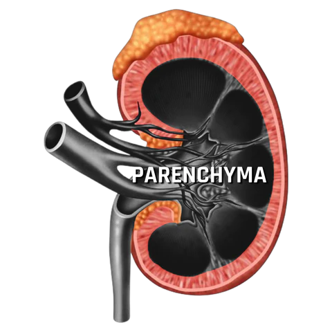
On the surface, the kidney appears to be highly complicated. However, there are two main parts when you break it down that can be impacted by Hydronephrosis. The most important part is the renal parenchyma which is the functional part of the kidney where urine is produced. The other is the pelvicalyceal system, also known as the calyceal system or “collecting system,” which collects and sends urine into the ureter.
The renal parenchyma is divided by two sub-organs, the medulla, and cortex. The pelvicalyceal system also has two sub-organs: the renal pelvis and calyces. The anatomy and physiology of these renal sub-organs are entirely different. As such, each part affects and behaves differently.
PELVICALYCEAL SYSTEM
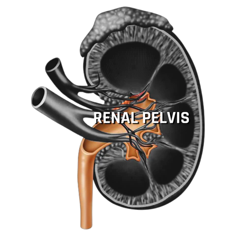
Renal Pelvis
The flexibility and expandability of the renal pelvis are high in most people. This is especially true in individuals with an extrarenal pelvic configuration compared to an intrarenal pelvic arrangement. Extrarenal pelvic configurations mean that more of the renal pelvis is located outside the kidney versus on the inside (see photo above).
The renal pelvis expands significantly to protect the renal parenchyma when faced with an obstruction leading to Hydronephrosis. This is true even in mild cases of Hydronephrosis where the increase in renal pelvic pressure is low. Because of this, the risk to the renal parenchyma (and your kidney function) is low.
However, in individuals with intrarenal pelvic configurations, the risk of renal damage can escalate quickly due to the low level of expansion available due to the more internal configuration of the renal pelvis.
Calyces
The expandability of the calyces is lower than that of the renal pelvis. Because of this, when the expansion of the calyces is observed during imaging, it represents a greater degree of Hydronephrosis (typically Grade 3 “Moderate” and higher) compared to an expansion of the renal pelvis alone (Grades 1 & 2 “Mild”). Like with the renal pelvis, the kidney calyces expand to protect the renal parenchyma.
RENAL PARENCHYMA
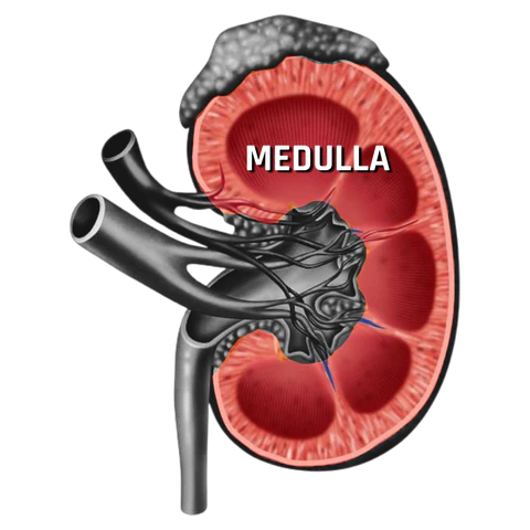
Medulla
The medulla is the interior of the kidney (see photo above), and its structure is somewhat similar to that of the lung. It is also the most expandable part of the renal parenchyma and can compress rapidly compared to the renal cortex. Depending on the degree of Hydronephrosis, the medulla becomes shorter and loses its pyramid form.
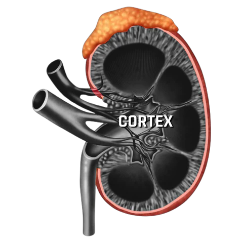
Cortex
The cortex is the exterior of the kidney (see photo above) and is the most important functional part. The average thickness of the context is > 6mm in healthy adults. The cortex is structurally similar to that of the liver, which is a relatively hard-solid organ. Because of this, any expansion (which leads to thinning) or contraction activity due to Hydronephrosis presents a significant risk of kidney damage.
In Grade 4 “Severe” Hydronephrosis, the differentiation between the cortex and the medulla is lost due to the over-expansion of the medulla. As a result, the thickness of the cortex decreases. When this grade of Hydronephrosis is identified through imaging, it is a very objective parameter. It is not operator dependent and is based on specific and measurable points. Additionally, it cannot be influenced by the level of hydration, bladder filling, position, or respiration.
To determine the thickness of the renal parenchyma, measurements are taken from the thinnest point on the longitudinal section of the kidney. Long-lasting thinning of the cortex is associated with low kidney function and a decrease in the number of nephrons. Because of this, a thinned cortex is suggestive of kidney damage. Furthermore, loss of more than half of the cortex (3mm+) is associated with kidney atrophy and irreversible kidney damage.
DEGREE OF HYDRONEPHROSIS
The quality of the renal parenchyma is based on thickness and appearance. Therefore, it is used to determine the severity of Hydronephrosis.
Thickness of Renal Parenchyma
Severe damage to the cortex and a decrease in the globular filtration rate (a measure of kidney health) occurs in individuals experiencing Grade 4 “Severe” Hydronephrosis. When it comes to prolonged exposure to this level of severe Hydronephrosis, the risk of permanent kidney damage is high (8-16%). Unfortunately, even after the blockage has been cleared, the function has been shown not to return to normal (thus why it is named permanent damage).
Appearance of the Renal Parenchyma
Many factors are assessed when determining damage based on the appearance of the renal parenchyma on Ultrasound. They are as follows:
- Hyperechogene parenchyma: a mild hepatic injury without significant vessel injury and indicates that surgical management of the hepatic injury is not required.
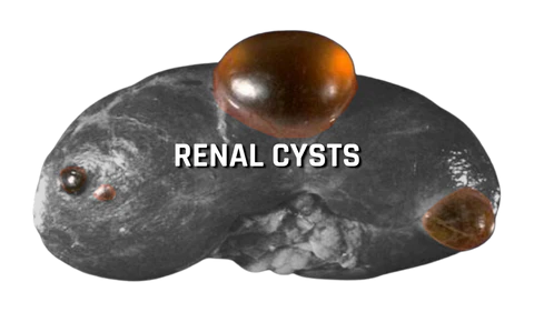
- Cystic degeneration in the cortex: the appearance of cysts in the cortex of the kidney suggesting repeated damage (see photo above).
- Loss of corticomedullary differentiation: occurs when the medulla and cortex become blended due to moderate or severe grades of Hydronephrosis.
CONCLUSIONS
We hope that this helps you better understand the mechanisms behind the risks of permanent kidney damage related to Hydronephrosis. As we covered in our blog/video on the Grades of Hydronephrosis, the risk of damage to the kidney only occurs in prolonged (48 hours or more) exposure to Grade 4 “Severe” Hydronephrosis. If this were to happen, you would be in so much extreme pain that you would have likely sought medical attention after a few hours of trying to endure this.

Written by Joey Weichmann
Joey Weichmann (aka “The Stone Slayer”) is the Founder of Stone Relief. As an avid naturopath and kidney stone sufferer, Joey has made it his mission to help health-conscious individuals stop kidney stones and get their life back.
Comments or questions?
Responses