Published: April 3, 2022 | 10 mins read
The Four Grades of Hydronephrosis [Will I Damage My Kidney?]
Hydronephrosis occurs when an obstruction leads to dilation (expansion) of the pelvicalyceal system (also known as the collecting system of the kidney) as a result of a back-up of urine into the kidney. Hydronephris can be caused by many other things and can be classified as either obstructive or non-obstructive. But, for today’s conversation, we will be focusing on obstructions caused by kidney stones.
- Pelvicalyceal System: formed by the renal pelvis and major/minor calyces inside the kidney.

SYMPTOMS
Hydronephrosis in and of itself is painless. However, when the back-up of urine into the kidney reaches a certain point (moderate to severe hydronephrosis), renal colic (the pain associated with kidney stones) becomes apparent. And, in some instances, VERY APPARENT. So much so that it oftentimes leads to 911 calls for an ambulance and a trip to the Emergency Room (ER).
However, hydronephrosis is a bit of a moving target. Individuals can be in the worst pain of their life at one moment, and be completely fine the next moment. It all has to do with the movement of your kidney stone through your urinary tract and any obstruction (mostly temporary) that it may cause along the way.

DIAGNOSIS
When it comes to identifying hydronephrosis, imaging is used just like when identifying your kidney stone. In fact, they are typically accomplished during the same imaging session. Currently, Computed Tomography (CT) is considered the gold standard for the diagnosis of kidney stones and renal colic as a result of hydronephrosis. As we have discussed in our blog on the Imaging Techniques for Kidney Stones, CT has sensitivities of 91-97% and specificities of 91-100% for detecting kidney stones, which are very high! Meaning that it is the most accurate option for identifying stones currently available.
- Sensitivity: Probability of a positive diagnostic test in a patient with the illness or injury.
- Specificity: Probability of a negative diagnostic test in a patient free of the disease or injury.
Beyond stone identification, Computed Tomography (CT) provides medical professionals with stone size, location, and density (attenuation), which can be helpful in predicting successful expulsive therapy versus the need for surgical intervention. However, as many as 50% of individuals diagnosed with kidney stones will have a recurrent episode and may receive multiple CT scans in their lifetime. This adds tremendous cost and exposure to radiation. As such, the role of Ultrasound (US) has gained popularity in recent years.
Ultrasound (US) has the advantage of using no radiation. However, the low sensitivity (72-87%) and specificity (73-83%) of Utlrasound (US) for identifying stone size and location may limit its usefulness in predicting the appropriate treatment plan and for follow-up with individuals experiencing renal colic.
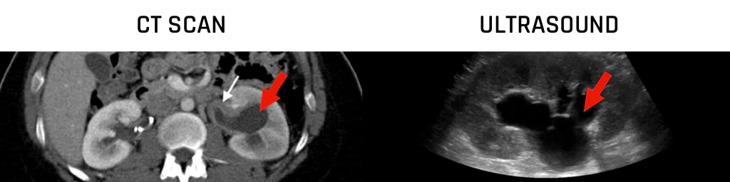
Another consideration to make is the ease of use for the technician and Urologist. As seen in the photo above, it is clear that the visibility of hydronephrosis is very clear on Computer Tomography (CT). But, a bit hazy on Ultrasound (US).
It is also important to note that the size of your stone does not always directly correlate with the grade of hydronephrosis. We have worked with tens of thousands of individuals and have seen stones as large as 11mm in the ureter not causing anything higher than mild hydronephrosis. Each individual will be presented with a situation that will be unique to their own. Do not let anyone convince you otherwise!
ONEN HYDRONEPHROSIS GRADING SYSTEM
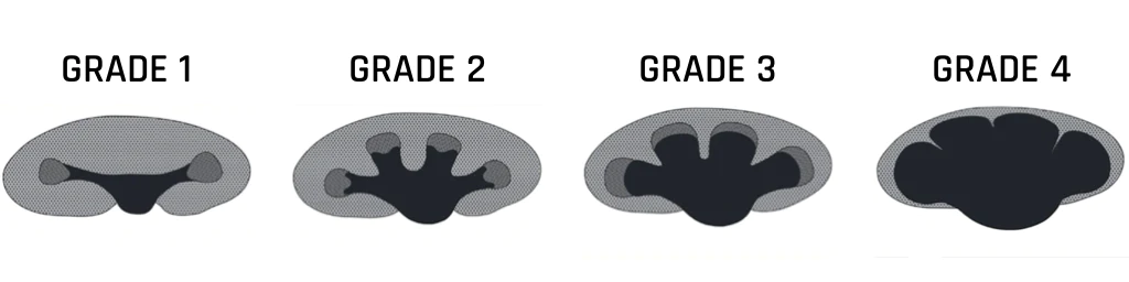
There are many different hydronephrosis grading systems in use around the world. Due to this, there is no current consensus on the “gold standard” grading system. To make things more complicated, many of the grading systems are highly variable and dependent on the skill of the Imaging Technician. However, there is one grading system that that has resolved all disadvantages of the other grading systems and promises accurate and easily reproducible grading results with a high degree of sensitivity and specificity, this is the Onen grading system.
The Onen grading system was developed for both adult and pediatric use, which has standardized the language Imaging Technicians and Urologists evaluate the kidneys. The terminology used is also the most clear out of all options. Therefore, all medical professionals interacting with you can speak the same language and be on the same page regarding treatment options.
The Onen grading system is evidence based and operates on reproducible parameters. In particular, there are two categories of kidney-related findings that are used when assigning a grade. The first is dilation of the pelvicalyceal system (i.e. how much urine is backing up into the kidney) and the second (and most important), the quality of the kidney parenchyma (made up of the medulla and cortex) in terms of thickness and quality.
This grading system divides thinning of the kidney parenchyma into two grades: medullary thinning and cortical thinning (see photo directly above). Quality is assessed on the appearance of the parenchyma. The presence of things like cysts can suggest kidney damage.
- NOTE: Thinning of the kidney parenchyma is what leads to the explosive pain experience in renal colic.

Grade 1 – Mild Hydronephrosis
Grade 1, also known as “mild” hydronephrosis will be present as a baseline for almost every kidney stone with very-very few exceptions. This is because the mere presence of a stone will have an impact on the flow of your urine. There’s no way around it (literally). As such, dilation is only observed in the renal pelvis (see photo above) and has not spread into the kidney itself.
However, it is important to note that this should not be feared! The impairment to your kidney function is limited and will return once the stone has passed. Do not let anyone scare you into a surgical procedure that you do not want and might not need out of fear.
Also important to note is that there is no pain associated with this grade of hydronephrosis as there is not any thinning of the kidney parenchyma. Thinning of the parenchyma is what associates with renal colic (kidney stone pain).
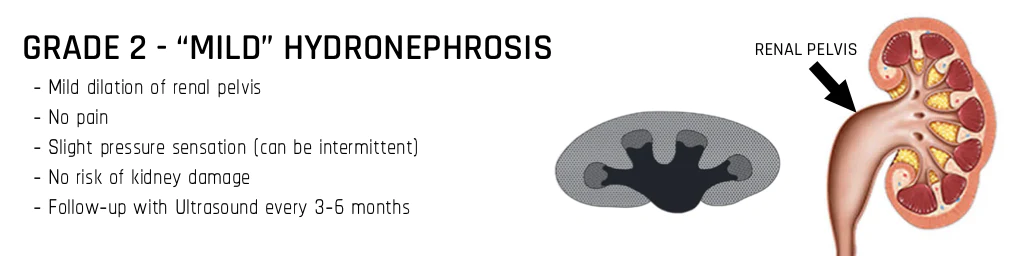
Grade 2 – Mild Hydronephrosis
In Grade 2, the calyceal system in addition to the renal pelvis become dilated due to urine back-up caused by your kidney stone. This is still considered to be “mild” hydronephrosis because there has been no change to the thickness of the kidney parenchyma (typically 10-14mm in adults). Remember, damage to the kidney and renal colic pain are associated with the thinning of the parenchyma.
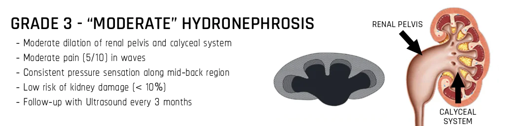
Grade 3 – Moderate Hydronephrosis
With Grade 3 hydronephrosis, things are starting to get a little more complicated. Both the renal pelvis and calyceal system are dilated just like within Grade 2, but now the total kidney parenchyma thickness (medulla and cortex) is starting to become thinned. In grade 3, the cortex remains normal. However, the medulla can reduce in thickness between 30-50%.
Pain will make it’s first appearance during Grade 3 hydronephrosis. While the pain will still fall short of the nuclear explosion level pain that likely signaled to you the presence of a problem (kidney stone). It will present itself as more of nagging ache with occasional spikes in pain through your day or week when the stone moves and creates a temporary obstruction that leads to the thinning of the parenchyma. The intervals of these spikes will vary from person to person.
In terms of damage to the kidney, the risks of permanent damage are still relatively low. However, it is important to listen to your body. If pain escalates past the point of your comfort, it may be time to seek medical attention to elevate the pain.
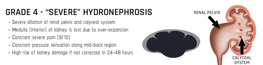
Grade 4 – Severe Hydronephrosis
Grade 4 hydronephrosis is likely something that you’ve already experienced for short spurts when you first learned of your kidney stone. This degree of obstruction is what leads to the nuclear reaction level pain that generally lands a majority of first-time kidney stone formers in the ER (or in an ambulance). And, while awful to experience, it is still an experience that you will want to file away for potential later use in identifying potentially serious problems while attempting to pass your stone.
In Grade 4 hydronephrosis, we are dealing with all of the issues from the previous Grades (dilation of the renal pelvis/calyceal system and thinning of the total kidney parenchyma). However, with severe hydronephrosis, the cortex becomes thin (< 3mm) and the medulla becomes a complete loss. Due to the stretching of the kidney that is occurring because of the urine back-up the recesses between the calyxes become significantly shorter and slim.
The loss of the medulla and the dramatic thinning of the cortex will prove to be very painful as mentioned above and should signal to you that it is time to seek medical attention. If left untreated, severe hydronephrosis can permanently damage your kidney. However, your body will give you ample signals and time to rectify the issue if you listen to the signs.

TREATMENT OPTIONS
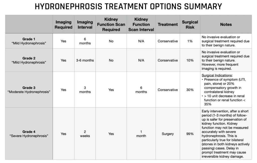
Grade 1 – Mild Hydronephrosis
Grade 1 cases neither require invasive evaluation, surgical treatment, or antibiotics due to their benign nature. All that is required is a follow-up with Ultrasound (US) to monitor the situation every 6 months.
Grade 2 – Mild Hydronephrosis
Grade 2 cases also neither require invasive evaluation, surgical treatment, or antibiotics due to their benign nature. And, just like with Grade 1, all that is required is a follow-up with Ultrasound (US) to monitor the situation every 3-6 months.
Grade 3 – Moderate Hydronephrosis
Grade 3 cases will require close follow-up including a renal scan to monitor for kidney functionality. But, outside of this, no surgery or antibiotics are required. However, a consistent Ultrasound evaluation of the state of your hydronephrosis should be set for roughly every 3 months until your stone has passed.
Grade 4 – Severe Hydronephrosis
Grade 4 cases will require surgical intervention to correct the obstruction. The surgical method chosen will vary from individual to individual based on Surgeon’s preference and the specific situation relating to your kidney stone. If you’d like to learn more about your surgical treatment options, please read our blog on the American Urological Association Guidelines.
It is important to note that with Grade 4 hydronephrosis a renal scan to assess kidney function is not possible. Surgery should be performed to remedy the obstruction as soon as possible and renal function assessed after 1 month. Timely and prompt surgical correction for sever hydronephrosis promises to improve any decreased kidney function.
CONCLUSION
Hydronephrosis can be a serious complication relating to kidney stones. However, it is a moving target and can change in minutes. When you arm yourself with the proper information, the fear that is typically instilled by medical professionals to spur you into surgery can be mitigated. Remember, at a baseline, if you have a kidney stone, you will have a presence of mild hydronephrosis. Your kidney functions may be slightly limited. But, this is not permanent and functionality will return over time. You won’t notice a thing.
However, if you are experiencing severe renal colic pain that lasts for more than 6 hours and cannot be remedied by physical activity to jostle the stone into a non-obstructing position, it is time to seek medical care to address the obstruction. Permanent damage can occur if not promptly treated. But again, this is only in instances of Grade 4 “Severe” hydronephrosis.
References

Written by Joey Weichmann
Joey Weichmann (aka “The Stone Slayer”) is the Founder of Stone Relief. As an avid naturopath and kidney stone sufferer, Joey has made it his mission to help health-conscious individuals stop kidney stones and get their life back.
Comments or questions?
Responses
WHAT TO READ NEXT
Publish Date: June 23, 2024
Do You Have a Phosphate Leak Leading to Kidney Stones?
Publish Date: June 9, 2024
The #1 Genetic Cause Of Kidney Stones: Cystinuria Disease
Publish Date: May 26, 2024
Distal Renal Tubular Acidosis Leads To Calcium Phosphate Stones