CALCIUM OXALATE KIDNEY STONES
Calcium Oxalate kidney stones are divided into two types by their predominant crystal structure. Type I are Calcium Oxalate Monohydrate (COM), and Type II is Calcium Oxalate Dihydrate (COD). Calcium oxalate Mono Hydrate (Type I) kidney stones present themselves in 5 unique forms (Type Ia-Ie). Calcium Oxalate Dihydrate (Type II) kidney stones present themselves in 3 distinct forms (Type IIa-IIc).
TYPE Ia COM STONES
Common Causes
Dietary hyperoxaluria, low duiresis (high oxalate conentration), or Randall's plaque
TYPE Ib COM STONES
Common Causes
Stasis, low diuresis, or total crystalline conversion from weddellite to whewellite
TYPE Id COM STONES
Common Causes
Malformative uropathy, stasis, and confined multiple stones
TYPE Ie COM STONES
Common Causes
Malformative uropathy, stasis, and confined multiple stones
TYPE IIa COD STONES
Common Causes
Hypercalciuria of various origin or a high molar ratio of calcium/citrate
TYPE IIb COD STONES
Common Causes
Hypercalciuria, hyperoxaluria, hypocitraturia, stasis, or low diuresis
TYPE IIc COD STONES
Common Causes
Hypercalciuria + malformative uropathy + stasis and confined multiple stones
MIXED COM & COD STONES
Common Causes
Various. Please click "Learn More" below for more information
TYPE I: CALCIUM OXALATE MONOHYDRATE (COM) KIDNEY STONES
The common cause for this type of kidney stone is an elevated concentration of oxalate ions in the urine without an associated elevation of urinary calcium. This excess concentration of oxalate ions is referred to as hyperoxaluria. Hyperoxaluria is caused by an inherited genetic disorder, intestinal disease, or by consuming too many oxalate-rich foods (Oxalate Content Food Guide). The specific subtype of these stones (TypeI-Ie) is determined by the level and duration of hyperoxaluria experienced by the individual.
MONOHYDRATE TYPE Ia STONES
Common Causes:
Hypercalciuria, hyperoxaluria, hypocitraturia, stasis, or low diuresis
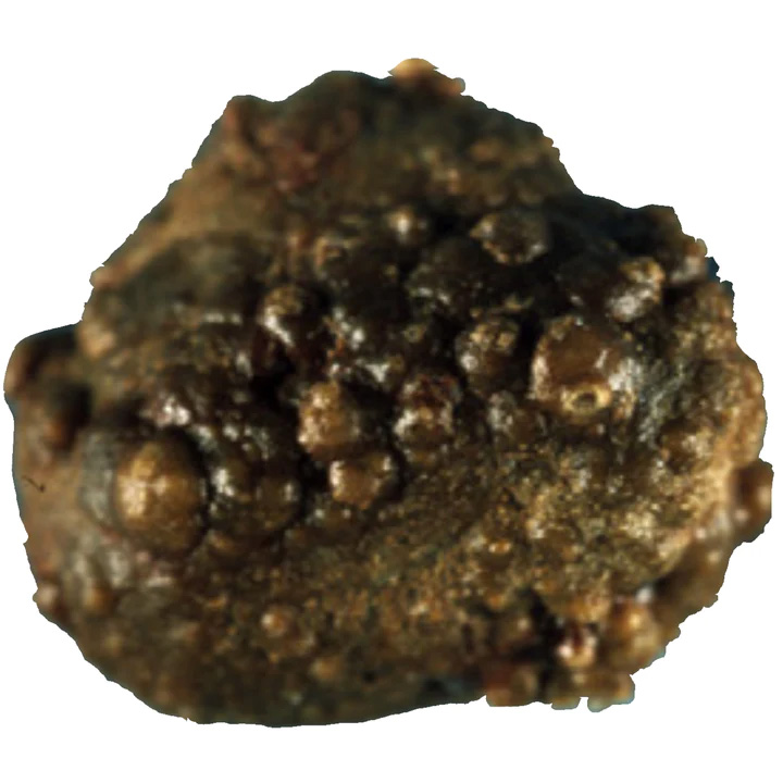
This type of stone is the most prevalent and is commonly referred to as an "idiopathic" calcium oxalate kidney stone. The idiopathic part refers to the stone being caused by external factors such as food/diet choices rather than genetic disorders/disease. Type Ia CaOx kidney stones represent 88% of Type I stones and 18% of all types of kidney stones.
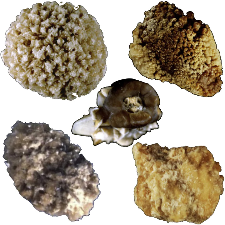
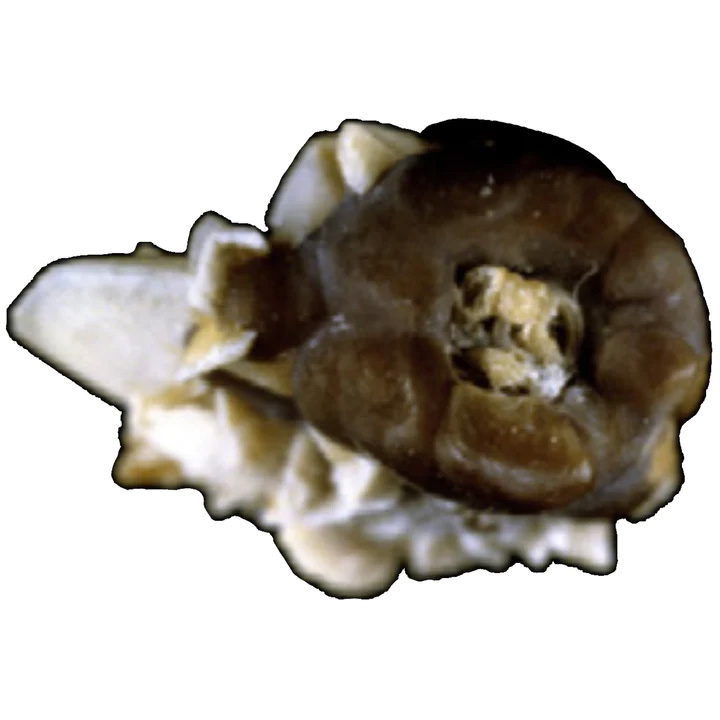
Their surface is typically spherical with a smooth, dark brown surface. When cut in half, the internal structure is composed of compact concentric layers with a radiating organization starting from the center. This internal structure is indicative of intermittent deposit of Calcium Oxalate Monohydrate crystals, coinciding with peaks of hyperoxaluria resulting from dietary habits.
Three conditions may be involved in the intermittent and moderate excessive concentration of oxalate in the urine. The most common cause is low water intake resulting in low urinary output. Other dietary factors may be:
High consumption of oxalate-rich foods like dark chocolate, beetroots, spinach, rhubarb, sorrel, star fruit, and many others (Oxalate Content Food Guide) High consumption of hydroxyproline-rich foods that induce a metabolic synthesis of oxalate by the liver. Low intake of calcium, resulting in increased absorption of oxalate ions by the gut.
Most Type Ia Calcium Oxalate Monohydrate kidney stones are dark brown. This color scheme is because of their intermittent growth, which lends itself to increased deposits of urinary pigments to be fixed on the crystals. In some rare cases, the surface of Type I CaOx stones exhibit a very thin and incomplete white layer made of very small Calcium Oxalate Monohydrate crystals. Because the underlying layers are dark brown, the thin superficial deposit appears as a grayish film.
This grayish film is significant as it points to a recent spike in hyperoxaluria, which leads to the new crystal deposits. Thus, dark brown Type Ia kidney stones are considered to be metabolically inactive. By contrast, grayish Type Ia kidney stones are metabolically active.
MONOHYDRATE TYPE Ib STONES
Common Causes:
Stasis, low diuresis, or total crystalline conversion from weddellite to whewellite
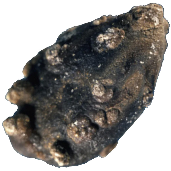
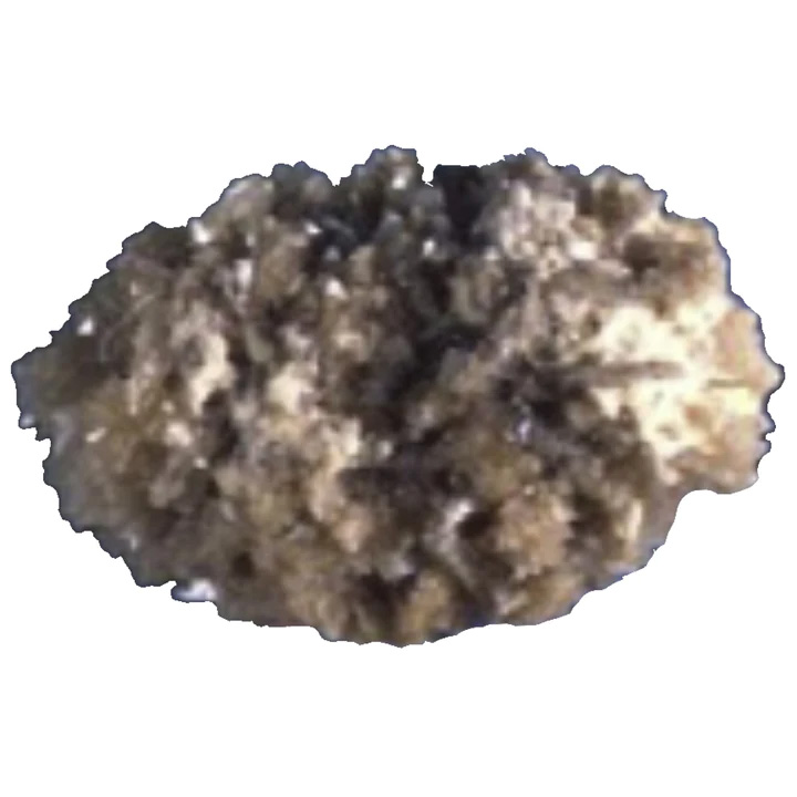
These types of kidney stones are also considered to be idiopathic. But, they are much less prevalent and represent only 5% of Type I Calcium Oxalate Monohydrate kidney stones. Type Ib kidney stones have a brown to dark brown oval shape with a rough surface. Their internal structure is also very unorganized. This structure lends itself to moderate hyperoxaluria.
MONOHYDRATE TYPE Ic STONES
Common Causes:
Primary hyperoxalurias (mainly type 1 by AGXT mutation)
Type Ic Calcium Oxalate Monohydrate kidney stones form because of Primary Hyperoxaluria Type 1 (PH1), the most severe form of nephrolithiasis. Primary Hyperoxaluria is a rare inherited genetic condition present at birth. In this condition, the liver does not create enough of a certain enzyme that regulates the production of oxalate.
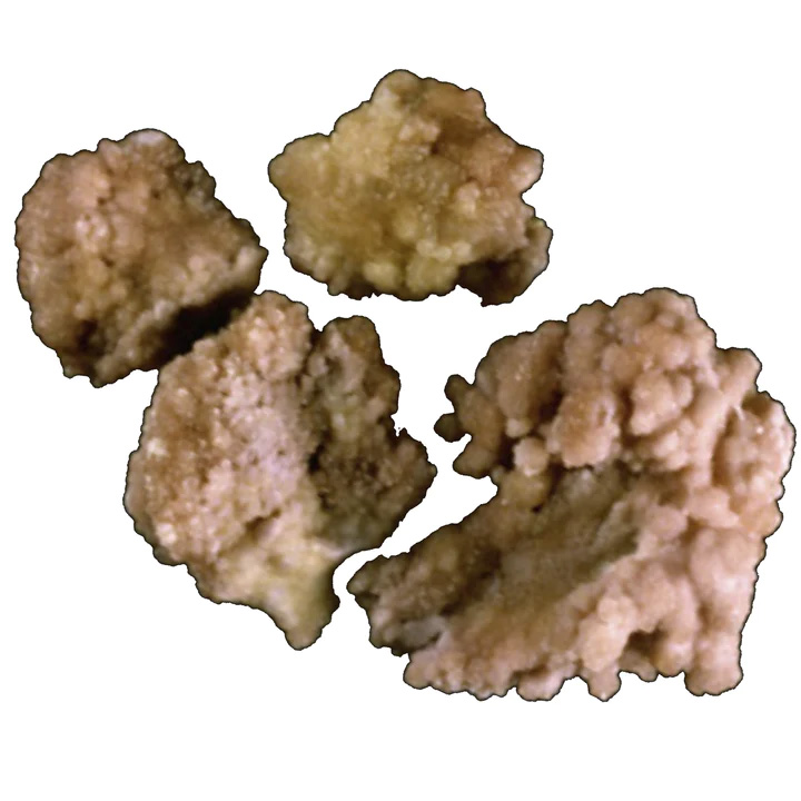
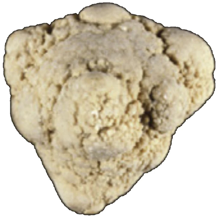
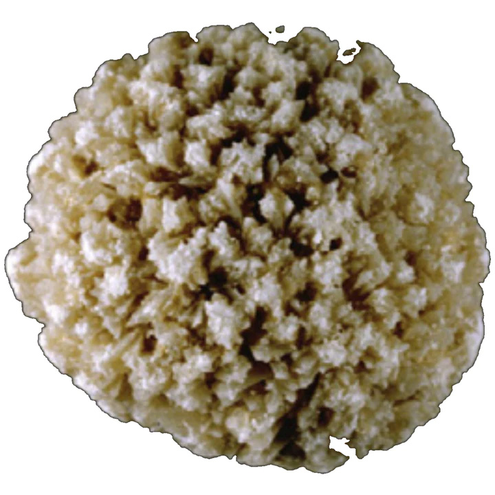
This type of kidney stone is made of pure calcium oxalate monohydrate. It exhibits a peculiar morphology of a pale-yellowish budding surface with an unorganized internal structure. Type Ic CaOx stones can also present themselves in a clear color, which should immediately alert doctors to the probability of Primary Hyperoxaluria Type 1 (PH1). Or, at least, of a condition associate with heavy hyperoxaluria.
Fortunately, Type Ic Calcium Oxalate Monohydrate kidney stones account for less than 1% of Type I kidney stones.
MONOHYDRATE TYPE Id STONES
Common Causes:
Malformative uropathy, stasis, and confined multiple stones
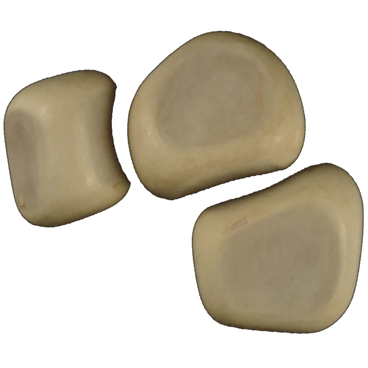
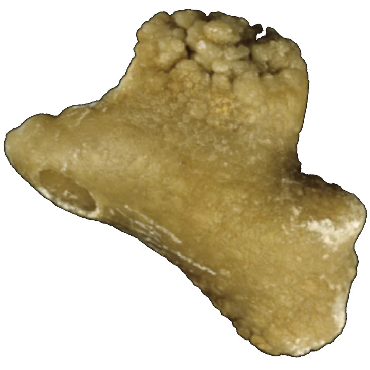
These types of kidney stones are also considered to be idiopathic. But, they are much less prevalent and represent only 4.1% of Type I Calcium Oxalate Monohydrate kidney stones. Type Id kidney stones present quite a different form factor with a very smooth surface. They are also beige or pale brown with an internal structure made up of thin concentric layers without a radiating pattern. These kidney stones are typically found in numbers within a person's kidneys; as they form in conditions of stasis within confined cavities such as diverticulum or hydronephrosis. The smooth surface is a result of the numerous stones rubbing against one another.
MONOHYDRATE TYPE Ie STONES
Common Causes:
Enteric hyperoxaluria, inflammatory bowel disease (Crohn's), illeal resections, and chronic pancreatitis
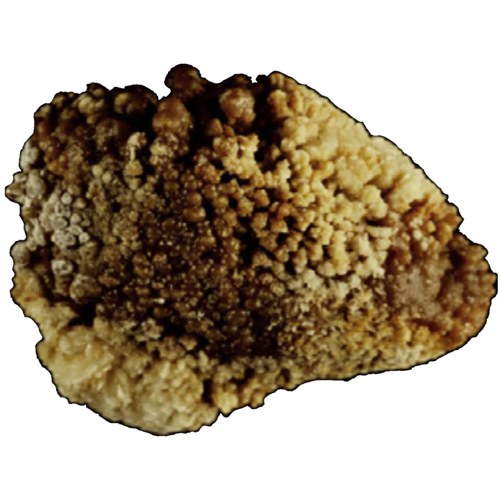
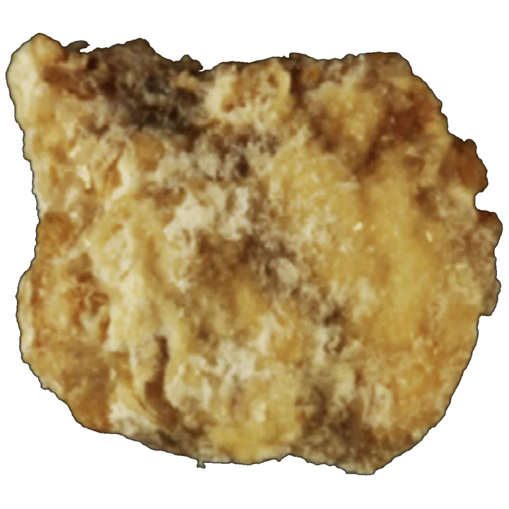
This type of COM stone is typically indicative of specific conditions associated with marked hyperoxaluria resulting from an intestinal dysfunction of diverse origins. This "Enteric Hyperoxaluria" is observed in inflammatory bowel disease (IBS/IBD). Enteric Hyperoxaluria is caused by several types of intestinal diseases, including Crohn's Disease and short-bowel syndrome as a result of surgical procedures, which can increase the amount of oxalate excreted in the urine. Type Ie kidney stones exhibit a locally budding, oval shape, with a rough pale brown-yellow surface. The internal structure can be best described as poorly organized dark brown layers with a radiating pattern of organization. Type Ie CaOx kidney stones represent 2% of Type I stones.
*NOTE: Calcium Oxalate Monohydrate kidney stones are not the unique type of stone that may develop in individuals with an inflammatory intestinal disease or after intestinal surgery [14]. Thus, it is crucial for people with these conditions to have their stone analyzed to identify the underlying stone-forming factors.
Another condition that is associated with the formation of Type Ie kidney stones is Type 2 diabetes. About 6% of kidney stones formed in diabetic individuals exhibit a form factor of Type Ie kidney stones. This correlation suggests that hyperoxaluria can be a metabolic disorder linked to Type 2 diabetes. Additionally, various metabolic pathways involving carbohydrates and polyols (low-digestible carbohydrates derived from the hydrogenation of their sugar or syrup source - e.g., lactitol from lactose) may be responsible for the formation of metabolic precursors of oxalate such as glyoxal or glyoxylate.
MONOHYDRATE TYPE Ie STONES
Common Causes:
Enteric hyperoxaluria, inflammatory bowel disease (Crohn's), illeal resections, and chronic pancreatitis
This type of COM stone is typically indicative of specific conditions associated with marked hyperoxaluria resulting from an intestinal dysfunction of diverse origins. This "Enteric Hyperoxaluria" is observed in inflammatory bowel disease (IBS/IBD). Enteric Hyperoxaluria is caused by several types of intestinal diseases, including Crohn's Disease and short-bowel syndrome as a result of surgical procedures, which can increase the amount of oxalate excreted in the urine. Type Ie kidney stones exhibit a locally budding, oval shape, with a rough pale brown-yellow surface. The internal structure can be best described as poorly organized dark brown layers with a radiating pattern of organization. Type Ie CaOx kidney stones represent 2% of Type I stones.
*NOTE: Calcium Oxalate Monohydrate kidney stones are not the unique type of stone that may develop in individuals with an inflammatory intestinal disease or after intestinal surgery [14]. Thus, it is crucial for people with these conditions to have their stone analyzed to identify the underlying stone-forming factors.
Another condition that is associated with the formation of Type Ie kidney stones is Type 2 diabetes. About 6% of kidney stones formed in diabetic individuals exhibit a form factor of Type Ie kidney stones. This correlation suggests that hyperoxaluria can be a metabolic disorder linked to Type 2 diabetes. Additionally, various metabolic pathways involving carbohydrates and polyols (low-digestible carbohydrates derived from the hydrogenation of their sugar or syrup source - e.g., lactitol from lactose) may be responsible for the formation of metabolic precursors of oxalate such as glyoxal or glyoxylate.
TYPE II: CALCIUM OXALATE DIHYDRATE (COD) KIDNEY STONES
This type of CaOx stone is commonly formed as a result of an elevated concentration of calcium ions in urine without a marked increase in urinary oxalate.
DIHYDRATE TYPE IIa STONES
Common Causes:
Hypercalciuria of various origin or a high molar ratio of calcium/citrate
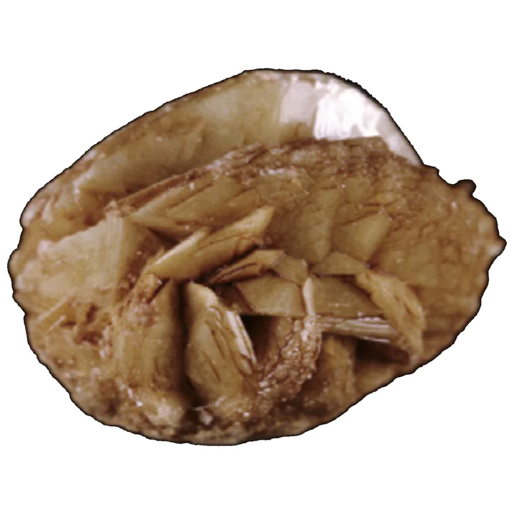
Type IIa kidney stones are the most commonly encountered and represent 69% of all Calcium Oxalate Dihydrate kidney stones. This type of stone has a yellow or light brown prickly-spiculated (lumpy and sharp) surface due to the presence of clustered bipyramidal crystals with sharp angles and edges. Their internal structure shows a loose radial crystallization.
Typically, this type of kidney stone is observed in individuals with idiopathic hypercalciuria. Hypercalciuria is an excess of calcium in the urine. It is caused by another condition, lending to high levels of calcium in the bloodstream. It is important to note that hypercalciuria is not related to dietary calcium intake.
DIHYDRATE TYPE IIb STONES
Common Causes:
Hypercalciuria, hyperoxaluria, hypocitraturia, stasis, or low diuresis
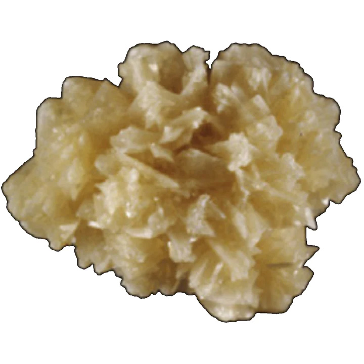
This type of COD kidney stone representation 30% of Type II stones. Type IIb kidney stones have a yellow or light brown surface with smooth-long bipyramidal crystals (often resembling small desert roses). Their internal structure is compact and poorly organized. This type of stone forms in individuals with both idiopathic hypercalciuria and moderate hyperoxaluria.
DIHYDRATE TYPE IIc STONES
Common Causes:
Hypercalciuria + malformative uropathy + stasis and confined multiple stones
Type IIc kidney stones are much less frequent and represent 0.7% of all COD stones. They have a rough gray-beige to pale-brown surface and a diffuse-concentric internal structure. This type of kidney stone is observed in individuals with both hypercalciuria and those with an unusual-obstructive urinary tract. These stones are typically found in numbers as a result of a stasis condition.
MIXED COM & COD KIDNEY STONES
Some Calcium Oxalate kidney stones present themselves as a mixture of Types Ia/IIa or Ia/IIb. These mixed stones are nearly as prevalent as pure CaOx stones. They account for 20.4% of all Calcium Oxalate type stones as compared to a prevalence of 29.4% for "pure" COM or COD kidney stones [19]. The composition can vary widely concerning percent COM versus COD.
Mixed CaOx kidney stones are observed in individuals with both hypercalciuria and hyperoxaluria, most often of dietary origin. Because the stone forming process is intermittent (not consistent), COD crystals may partly convert to COM (COM is the thermodynamically stable phase of calcium oxalate) [20]. As a result of the mixture of COM and COD, stones often present themselves with many of the typical traits of COD crystals overlapped by COM crystals.
TYPE IIA + TYPE Ia/IIb + TYPE IVa
Common Causes
Hyperoxaluria + hypercalciuria
TYPE 1a/IIA + TYPE Ib/IIB + TYPE IVa/IIb
Common Causes
Intermittent or moderate hypercalciuria, crystalline conversion, moderately high oxalate concentration, or low diuresis
TYPE Ia/IIb + TYPE Ia + TYPE IVa
Common Causes
Moderately high oxalate concentration, low diuresis, intermittent hypercalciuria, or Vitamin D hypersensitivity
TYPE IIa/IVa1
Common Causes
Hypercalciuria
TYPE IIa/IVa1
Common Causes
Resorptive or absorptive hypercalciuria or primary hyperparathyroidism
CALCIUM OXALATE TRIHYDRATE (COT) OR CAOXITE KIDNEY STONES
Caoxite is the third crystalline form of calcium oxalate identified in urinary stones and crystals [21]. Like COM kidney stones, this type of stone is associate with hyperoxaluric states. However, given that this is a rare stone type, it suggests that other factors are involved in its formation.
Caoxite has been observed in two very different situations: 1) CaOx stones induced by a drug prescribed for cardiovascular diseases [22] and 2) CaOx stones formed in patients with primary hyperoxaluria.
In both cases, COT was mixed with COM. From a thermodynamic point of view, calcite is a volatile form of calcium oxalate which spontaneously and rapidly converts to COM. In in vitro studies, caoxite was shown to transform into COM within a few hours. The presence of caoxite in kidney stones suggests that it is stabilized in the urine of affected individuals. Caoxite crystals are believed to be caused by an imbalance between the promoters of calcium oxalate formation and macromolecules in the urine.
CLOSING NOTES ON CAOX STONE FORMERS
Among common CaOx stones, the predominance of COD Type IIa suggests hypercalciuria (excess calcium in the urine) as the main contributing factor. And, Type IIb is suggestive of hypercalciuria associated with moderate hyperoxaluria (excessive urinary excretion of oxalate). The predominance of COM Type Ia is most often associated with an increased urine oxalate concentration resulting from inadequate liquid intake and moderately increased oxalate excretion, often of dietary origin.